Скачать с ютуб Epidural Hemorrhage | Anatomical | Features & Pathophysiology | Dr Najeeb в хорошем качестве
Epidural Hemorrhage | Anatomical | Features & Pathophysiology | Dr Najeeb
2 года назад
Скачать бесплатно Epidural Hemorrhage | Anatomical | Features & Pathophysiology | Dr Najeeb в качестве 4к (2к / 1080p)
У нас вы можете посмотреть бесплатно Epidural Hemorrhage | Anatomical | Features & Pathophysiology | Dr Najeeb или скачать в максимальном доступном качестве, которое было загружено на ютуб. Для скачивания выберите вариант из формы ниже:
Загрузить музыку / рингтон Epidural Hemorrhage | Anatomical | Features & Pathophysiology | Dr Najeeb в формате MP3:
Если кнопки скачивания не
загрузились
НАЖМИТЕ ЗДЕСЬ или обновите страницу
Если возникают проблемы со скачиванием, пожалуйста напишите в поддержку по адресу внизу
страницы.
Спасибо за использование сервиса savevideohd.ru
Epidural Hemorrhage | Anatomical | Features & Pathophysiology | Dr Najeeb
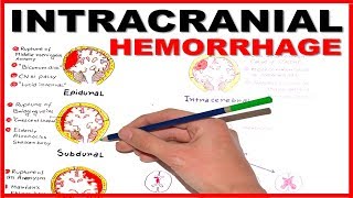
#Epiduralheamorrhage #Anatomicalcorrelation #drnajeeblectures Epidural Hemorrhage | Anatomical Correlation Clinical | Features & Pathophysiology | Dr Najeeb Like this video? Sign up now on our website at https://www.DrNajeebLectures.com to access 800+ Exclusive videos on Basic Medical Sciences & Clinical Medicine. These are premium videos (NOT FROM YOUTUBE). All these videos come with English subtitles & download options. Sign up now! Get Lifetime Access for a one-time payment of $45 ONLY! Sign up now on our website at https://members.drnajeeblectures.com/ --------------------------------------------------------------------------------------------------------------------------- Why sign up for premium membership? Here's why! Membership Features for premium website members. 1. More than 800+ Medical Lectures. 2. Basic Medical Sciences & Clinical Medicine. 3. Mobile-friendly interface with android and iOS apps. 4. English subtitles and new videos every week. 5. Download option for offline video playback. 6. Fanatic customer support and that's 24/7. 7. Fast video playback option to learn faster. 8. Trusted by over 2M+ students in 190 countries. --------------------------------------------------------------------------------------------------------------------------- ▬▬▬▬▬▬▬▬▬▬ Contents of this video ▬▬▬▬▬▬▬▬▬▬ Epidural Hemorrhage Anatomical Correlation Clinical Features And Pathophysiology Epidural hemorrhage or Extradural hemorrhage occurs when there is hemorrhage in the extradural space. Extradural space lies between the periosteal layer and meningeal layer of the dura matter. These two layers are very close to each other but there is potential space between them. These layers are held tightly together at skull sutures. In some places, these two layers separate making venous dural sinuses. Anterior branch of middle meningeal artery and vein runs between two layers of dura matter. When there is rupture of anterior branches of middle meningeal artery or vein due to trauma, epidural hematoma occurs and expands between skull bone and meningeal layer of dura making a biconvex shape or crescent shape hematoma. Hematoma does not expand beyond skull sutures Clinically epidural hematoma presents with 'Lucid interval' Lucid interval is a phenomenon in which a person initially loses consciousness after trauma to the head but immediately regains consciousness and then there is progressive loss of consciousness. Ct scan is the modality of choice in diagnosing epidural hemorrhage. Surgical intervention (burr hole craniotomy) is the treatment of choice. Join this channel to get access to perks: Sign up now on our website at https://members.drnajeeblectures.com/... / @doctornajeeb Follow us on Facebook :- / drnajeeb Follow us on Instagram :- / drnajeeblectures
Comments


![Subdural vs Epidural Hematoma/Hemorrhage [CT Scan Findings]](https://i.ytimg.com/vi/KW_kD_wGAUM/mqdefault.jpg)