РЎРәР°СҮР°СӮСҢ СҒ СҺСӮСғРұ Animated Gross anatomy of Appendix: Position, Blood supply, Venous drainage, Nerve supply, Histology РІ С…РҫСҖРҫСҲРөРј РәР°СҮРөСҒСӮРІРө
Animated Gross anatomy of Appendix: Position, Blood supply, Venous drainage, Nerve supply, Histology
6 Р»РөСӮ РҪазаРҙ
РЎРәР°СҮР°СӮСҢ РұРөСҒРҝлаСӮРҪРҫ Animated Gross anatomy of Appendix: Position, Blood supply, Venous drainage, Nerve supply, Histology РІ РәР°СҮРөСҒСӮРІРө 4Рә (2Рә / 1080p)
РЈ РҪР°СҒ РІСӢ РјРҫР¶РөСӮРө РҝРҫСҒРјРҫСӮСҖРөСӮСҢ РұРөСҒРҝлаСӮРҪРҫ Animated Gross anatomy of Appendix: Position, Blood supply, Venous drainage, Nerve supply, Histology или СҒРәР°СҮР°СӮСҢ РІ РјР°РәСҒималСҢРҪРҫРј РҙРҫСҒСӮСғРҝРҪРҫРј РәР°СҮРөСҒСӮРІРө, РәРҫСӮРҫСҖРҫРө РұСӢР»Рҫ загСҖСғР¶РөРҪРҫ РҪР° СҺСӮСғРұ. ДлСҸ СҒРәР°СҮРёРІР°РҪРёСҸ РІСӢРұРөСҖРёСӮРө РІР°СҖРёР°РҪСӮ РёР· С„РҫСҖРјСӢ РҪРёР¶Рө:
ЗагСҖСғР·РёСӮСҢ РјСғР·СӢРәСғ / СҖРёРҪРіСӮРҫРҪ Animated Gross anatomy of Appendix: Position, Blood supply, Venous drainage, Nerve supply, Histology РІ С„РҫСҖРјР°СӮРө MP3:
Р•СҒли РәРҪРҫРҝРәРё СҒРәР°СҮРёРІР°РҪРёСҸ РҪРө
загСҖСғзилиСҒСҢ
РқРҗР–РңРҳРўР• ЗДЕСЬ или РҫРұРҪРҫРІРёСӮРө СҒСӮСҖР°РҪРёСҶСғ
Р•СҒли РІРҫР·РҪРёРәР°СҺСӮ РҝСҖРҫРұР»РөРјСӢ СҒРҫ СҒРәР°СҮРёРІР°РҪРёРөРј, РҝРҫжалСғР№СҒСӮР° РҪР°РҝРёСҲРёСӮРө РІ РҝРҫРҙРҙРөСҖР¶РәСғ РҝРҫ Р°РҙСҖРөСҒСғ РІРҪРёР·Сғ
СҒСӮСҖР°РҪРёСҶСӢ.
РЎРҝР°СҒРёРұРҫ Р·Р° РёСҒРҝРҫР»СҢР·РҫРІР°РҪРёРө СҒРөСҖРІРёСҒР° savevideohd.ru
Animated Gross anatomy of Appendix: Position, Blood supply, Venous drainage, Nerve supply, Histology
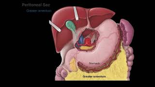
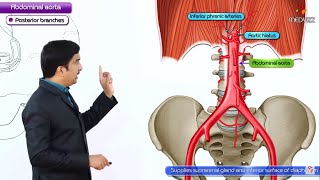
рҹ“Ң рқҗ…рқҗЁрқҗҘрқҗҘрқҗЁрқҗ° рқҗЁрқҗ§ рқҗҲрқҗ§рқҗ¬рқҗӯрқҗҡрқҗ рқҗ«рқҗҡрқҗҰ:- В В /В drgbhanuprakashВ В рҹ“Ңрқ—қрқ—јрқ—¶рқ—» рқ—ўрқҳӮрқ—ҝ рқ—§рқ—Ірқ—№рқ—Ірқ—ҙрқ—ҝрқ—®рқ—ә рқ—–рқ—өрқ—®рқ—»рқ—»рқ—Ірқ—№ рқ—ӣрқ—Ірқ—ҝрқ—І:- https://t.me/bhanuprakashdr рҹ“Ңрқ—ҰрқҳӮрқ—ҜрқҳҖрқ—°рқ—ҝрқ—¶рқ—Ҝрқ—І рқ—§рқ—ј рқ— рқҳҶ рқ— рқ—®рқ—¶рқ—№рқ—¶рқ—»рқ—ҙ рқ—ҹрқ—¶рқҳҖрқҳҒ:- https://linktr.ee/DrGBhanuprakash Animated Gross anatomy of Appendix - Position, Blood supply, Venous drainage, Nerve supply and Histology The cecum and appendix are part of the gastrointestinal tract. They are the most proximal part of the large intestine, located between the ileum (distal small bowel) and the ascending colon. Having served as a site for cellulose digestion in our ancestors, the cecum now simply acts as a reservoir for chyme which it receives from the ileum. The appendix contains a large amount of lymphoid tissue, but has no vital functions in the human. In this article, we shall look at the anatomy of the cecum and appendix вҖ“ their anatomical structure and relations, neurovascular supply and lymphatic drainage. Anatomical Structure and Relations The cecum. Note the blind end inferiorly, and its continuity with the ascending colon superiorly. The cecum. Note the blind end inferiorly, and its continuity with the ascending colon superiorly. The cecum is the most proximal part of the large intestine and can be found in the right iliac fossa and suprapubic region. It lies slightly inferior to the iliocaecal junction, and can be palpated in the right iliac fossa if enlarged due to faeces, gas or malignancy. The name is derived from the Latin word вҖҳcaecusвҖҷ, meaning вҖҳblindвҖҷ, due to its blind-end inferiorly. Superiorly, however, the open end of the cecum is continuous with the ascending colon. Unlike the ascending colon above it, the cecum is intraperitoneal. The appendix, also known as the vermiform (worm-shaped) appendix is a narrow, blind ended tube attached to the posteromedial end of the cecum. The position of the free-end of the appendix is highly variable, and can be categorised into seven main locations. The most common positions are retrocaecal and subileal. A simple way to remember the positions is by imagining the appendix as the hour hand of a clock: Pre-ileal вҖ“ Anterior to the terminal ileum вҖ“ 1 oвҖҷclock. Post-ileal вҖ“ Posterior to the terminal ileum вҖ“ 2 oвҖҷclock. Sub-ileal вҖ“ Parallel with the terminal ileum вҖ“ 3 oвҖҷclock. Pelvic вҖ“ Descending over the pelvic brim вҖ“ 5 oвҖҷclock. Sub-caecal вҖ“ Below the cecum вҖ“ 6 oвҖҷclock. Paracaecal вҖ“ Alongside the lateral border of the cecum вҖ“ 10 oвҖҷclock. Retrocaecal вҖ“ Behind the cecum вҖ“ 11 oвҖҷclock. Neurovascular Supply ----------------------------------- The cecum is derived from the embryologic midgut вҖ“ and therefore blood is supplied by a branch of the superior mesenteric artery. The branch is the ileocolic artery, which splits into anterior and posterior caecal arteries to supply the cecum. Venous drainage is provided by the ileocolic vein, which empties into the superior mesenteric vein. The appendix receives its blood supply via the appendicular artery (derived from the ileocolic artery), and drains through the appendicular vein. Both are contained, along with lymphatic vessels and nerves, within the mesoappendix, a fold of mesentery which suspends the appendix from the terminal ileum. The autonomic nervous system innervates the cecum and appendix. It achieves this by means of the ileocolic branch of the superior mesenteric plexus, which follows the same course as the ileocolic artery. The cecum, appendix and ascending colon. Note the inferior position of the cecum in relation to the ileum. Arterial supply to the cecum and appendix, via the ileocecal artery. Lymphatic Drainage ------------------------------- Lymph from the cecum and appendix ultimately drains into the upper and lower ileocolic lymph nodes, which surround the ileocolic artery. However, lymph from the cecum travels via a number of intermediate mesenteric nodes, whereas that of the appendix travels via a single intermediate node in the mesoappendix. Clinical Relevance: Appendicitis --------------------------------------------------- Appendicitis is acute inflammation of the appendix, and is the most common cause for acute, severe abdominal pain. The abdomen is most tender at McBurneyвҖҷs point вҖ“ one third of the distance from the right anterior superior iliac spine to the umbilicus. This corresponds to the location of the base of the appendix. The aetiology depends on age. In the young, it is mostly due to an increase in lymphoid tissue size, which occludes the lumen. From 30 years old onwards, it is more likely to be blocked due to faecal matter. Initially, the appendix cannot drain, and so increases in size, stretching the visceral peritoneum. This causes a vague pain in the periumbilical region. As the appendix swells, it irritates the parietal peritoneum, and causes severe pain in the right lower quadrant. If the appendix is not removed, it can become necrotic and rupture, resulting in peritonitis (inflammation of the peritoneum).
Comments