Скачать с ютуб Anatomy Of The Psoas & Iliacus Muscles - Everything You Need To Know - Dr. Nabil Ebraheim в хорошем качестве
Anatomy Of The Psoas & Iliacus Muscles - Everything You Need To Know - Dr. Nabil Ebraheim
9 лет назад
Из-за периодической блокировки нашего сайта РКН сервисами, просим воспользоваться резервным адресом:
Загрузить через ClipSave.ruСкачать бесплатно Anatomy Of The Psoas & Iliacus Muscles - Everything You Need To Know - Dr. Nabil Ebraheim в качестве 4к (2к / 1080p)
У нас вы можете посмотреть бесплатно Anatomy Of The Psoas & Iliacus Muscles - Everything You Need To Know - Dr. Nabil Ebraheim или скачать в максимальном доступном качестве, которое было загружено на ютуб. Для скачивания выберите вариант из формы ниже:
Загрузить музыку / рингтон Anatomy Of The Psoas & Iliacus Muscles - Everything You Need To Know - Dr. Nabil Ebraheim в формате MP3:
Если кнопки скачивания не
загрузились
НАЖМИТЕ ЗДЕСЬ или обновите страницу
Если возникают проблемы со скачиванием, пожалуйста напишите в поддержку по адресу внизу
страницы.
Спасибо за использование сервиса savevideohd.ru
Anatomy Of The Psoas & Iliacus Muscles - Everything You Need To Know - Dr. Nabil Ebraheim
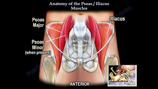
Dr. Ebraheim’s educational animated video describes the anatomy of the sartorius muscle. Psoas major origin: the psoas major muscle arises from the transverse processes and the lateral aspects of the vertebral bodies T12-L5. Psoas major insertion: the psoas runs downward across the pelvic brim and then passes deep to the ilioinguinal ligament where it then forms a tendon past the hip joint capsule which inserts into the lesser trochanter of the femur. Psoas major innervation: innervation of the psoas major occurs from L1 to L3 of the lumbar plexus. (L1,L2,L3). Psoas major function: hip flexion. Iliacus origin: the iliacus arises from the iliac fossa on the interior side of the hip bone and also from the region of the anterior inferior iliac spine (AIIS). Illiacus insertion: the iliacus inserts into the base of the lesser trochanter of femur. Innervation: the iliopsoas is innervated by the femoral nerve and direct branches from the lumbar plexus. Iliacus function: as a part of the iliopsoas, the iliacus contributes to flexion of the hip joint. The iliopsoas tendon is separated from the hip joint capsule by the iliopsoas bursa. Psoas and iliacus muscle assessment Testing the hip flexion strength: as the patient lifts the knee straight up off the examination table, the examiner then applies download pressure onto the knee to in order assess the hip flexion strength. Iliopsoas compartment syndrome: Both muscles are in the extraperitoneal space, or referred to as the iliopsoas compartment. Muscles within the compartment: iliacus, psoas major, psoas minor (when present). Causes of compartment syndrome in the pelvis: the pelvis is extremely rare area for compartment syndrome to develop. However, hemorrhage in the pelvis and iliopsoas hematoma may usually be caused by: severe trauma, anti-coagulation therapy, hemophilia or other blood diseases. Clinical presentation: flexion attitude of the involved hip. Pain with passive extension of the involved hip. Tenderness along the inguinal ligament. Paresthesia around the medial side of the knee in the distribution of the saphenous nerve. Diagnosis and treatment of iliopsoas hematoma: •Measurement of pressure is difficult. •MRI or CT scan for diagnosis. •Conservative treatment with observation. •Correction of coagulation deficit if applicable. •Surgical intervention is rarely required. Iliospsoas abscess: a primary abscess is caused by hematogenous spread of infection. The infection starts in the muscle itself. In a secondary abscess, the infection spreads from another area to the psoas muscle. For example, the infection may travel from the spine when it is infected by tuberculosis (Pott’s disease). Historically this is the cause of the psoas abscess. It can also spread from the SI joint, kidneys or bowels. The iliopsoas abscess may initially present with signs and symptoms in the buttock, hip or thigh. Such signs and symptoms may be obscure, non-specific and misleading. Abscess of the iliopsoas muscle is a diagnostic dilemma with a difficult diagnosis that is often delayed. The patient may be lying supine with the hip flexed and refuses to move, resisting any attempt for examination. With psoas involvement, the hip appears to be flexed, with limited and painful range of motion. This diverts attention away from the abdomen or pelvic source of the abscess. The patient may have a low-grade fever and cannot straighten the leg. A high index of suspicion is necessary and diagnosis is aided by performing the psoas sign. The psoas sign is helpful in diagnosing a psoas abscess. The patient is positioned on the side and the hip is extended to see if there is pain present in the iliopsoas region. Become a friend on facebook: / drebraheim Follow me on twitter: https://twitter.com/#!/DrEbraheim_UTMC Donate to the University of Toledo Foundation Department of Orthopaedic Surgery Endowed Chair Fund: https://www.utfoundation.org/foundati... Background music provided as a free download from YouTube Audio Library. Song Title: Every Step
Comments