Скачать с ютуб Melanocytic Dermpath Basics: Melanoma в хорошем качестве
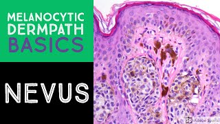
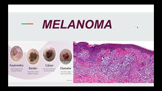
Melanocytic Dermpath Basics: Melanoma
6 лет назад
Скачать бесплатно Melanocytic Dermpath Basics: Melanoma в качестве 4к (2к / 1080p)
У нас вы можете посмотреть бесплатно Melanocytic Dermpath Basics: Melanoma или скачать в максимальном доступном качестве, которое было загружено на ютуб. Для скачивания выберите вариант из формы ниже:
Загрузить музыку / рингтон Melanocytic Dermpath Basics: Melanoma в формате MP3:
Если кнопки скачивания не
загрузились
НАЖМИТЕ ЗДЕСЬ или обновите страницу
Если возникают проблемы со скачиванием, пожалуйста напишите в поддержку по адресу внизу
страницы.
Спасибо за использование сервиса savevideohd.ru
Melanocytic Dermpath Basics: Melanoma
A complete organized library of all my videos, digital slides, pics, & sample pathology reports is available here: https://kikoxp.com/posts/5084 (dermpath) & https://kikoxp.com/posts/5083 (bone/soft tissue sarcoma pathology). Part 2 of my (in progress) video series about the pathology of melanocytic skin lesions. This video discusses the basic features and patterns that are commonly seen in melanoma, including asymmetry, pagetoid spread, confluent growth, abnormal/lack of maturation, severe cytologic atypia, and mitotic activity in the invasive dermal component. These are not hard and fast rules, just a helpful framework to serve as a starting point for understanding how to diagnose melanocytic lesions histologically. Exceptions exist for pretty much all of these things. Watch the video to learn more. Many thanks to my awesome medical student, Gray Orman, for transcribing this entire video so that closed captions would be available for viewers who need them! You can download the entire transcript of the video as a .docx here: http://bit.ly/2ZhUyMa. Here's an excellent example of confluent growth in an acral lentiginous melanoma (WSI digital whole slide image): https://kikoxp.com/posts/2813/. And here's a video explaining the case in more detail: https://kikoxp.com/posts/2836/. Topics Discussed (click timestamp to jump to that part of the video): Asymmetry- 6:40 Pagetoid Spread- 7:30 Severe Cytologic Atypia- 11:15 Mitotic Activity in Dermal Component- 16:00 and 1:07:50 Abnormal/Lack of Maturation- 21:53 and 1:09:40 Confluent Growth- 32:55 Breslow Depth- 49:05 Regression- 1:00:10 Ulceration- 1:03:20 Pattern of Metastatic or Recurrent Melanoma- 1:10:05 Melanoma subtypes: Superficial Spreading- 29:10 (and before) Acral Lentiginous- 30:24 Unzipping Sign (Melanocytic Blistering)- 37:30 Eccrine Duct Wrapping- 39:30 Lentigo maligna- 50:30 Immunostaining- 52:30 Nodular- 1:03:10 Other videos that will help you better understand this topic: -Normal Skin Histology: • Normal Skin Histology - Explained by ... -Nevus Basics: • Melanocytic Dermpath Basics: Benign N... -Melanocytic Immunohistochemistry: • Melanocytic Dermpath Basics: Immunohi... Disclaimers: As I said in the video, distinguishing nevus from melanoma is serious business. My video does not replace the need for getting an expert consultation should you encounter a difficult melanocytic lesion in your practice. Also, melanocytic dermpath has areas of controversy with differing strongly held points of view. by various experts in the field. This video represents MY current views as of May 23, 2018. My views have changed since I started practice, and I suspect (and hope) that they will continue to evolve over time. You (or your mentors) may do things differently than I do. That's ok by me. I'm just sharing the way I currently think of melanocytic lesions with the hope that viewers may find it useful. My goal is to educate, not to create dogma. This video is geared towards medical students, pathology or dermatology residents, or practicing pathologists or dermatologists. Of course, this video is for educational purposes only and is not formal medical advice or consultation. Presented by Jerad M. Gardner, MD. Please subscribe to my channel to be notified of new pathology teaching videos. Follow me on: Snapchat: JMGardnerMD Twitter: @JMGardnerMD Instagram: @JMGardnerMD Facebook: / jmgardnermd
Comments