Скачать с ютуб Lecture: How to Interpret OCT Findings in the Diagnostic Evaluation of Glaucoma в хорошем качестве
Lecture: How to Interpret OCT Findings in the Diagnostic Evaluation of Glaucoma
2 года назад
Скачать бесплатно Lecture: How to Interpret OCT Findings in the Diagnostic Evaluation of Glaucoma в качестве 4к (2к / 1080p)
У нас вы можете посмотреть бесплатно Lecture: How to Interpret OCT Findings in the Diagnostic Evaluation of Glaucoma или скачать в максимальном доступном качестве, которое было загружено на ютуб. Для скачивания выберите вариант из формы ниже:
Загрузить музыку / рингтон Lecture: How to Interpret OCT Findings in the Diagnostic Evaluation of Glaucoma в формате MP3:
Если кнопки скачивания не
загрузились
НАЖМИТЕ ЗДЕСЬ или обновите страницу
Если возникают проблемы со скачиванием, пожалуйста напишите в поддержку по адресу внизу
страницы.
Спасибо за использование сервиса savevideohd.ru
Lecture: How to Interpret OCT Findings in the Diagnostic Evaluation of Glaucoma
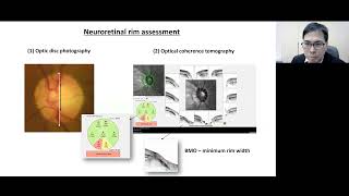
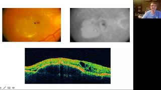
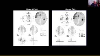
Optical coherence tomography (OCT) has become an indispensable tool in the diagnostic evaluation of glaucoma. However, deciding what regions (e.g., optic disc region vs. macula) and what parameters (e.g., retinal nerve fiber layer (RNFL) thickness vs. ganglion cell inner plexiform layer (GCIPL) thickness vs. neuroretinal rim width) to measure can often be confusing. False positives and false negatives in OCT reports are common. This webinar will walk through examples from different OCT models and elaborate correct interpretation of OCT findings to aid diagnosis and monitoring of glaucoma. Lecturer: Dr. Chris Leung, Head of Dept., Clinical Professor, Dept. of Ophthalmology, The University of Hong Kong
Comments