Скачать с ютуб Osteochondroma , solitary and multiple . Everything You Need To Know - Dr. Nabil Ebraheim в хорошем качестве
Osteochondroma , solitary and multiple . Everything You Need To Know - Dr. Nabil Ebraheim
8 лет назад
Скачать бесплатно Osteochondroma , solitary and multiple . Everything You Need To Know - Dr. Nabil Ebraheim в качестве 4к (2к / 1080p)
У нас вы можете посмотреть бесплатно Osteochondroma , solitary and multiple . Everything You Need To Know - Dr. Nabil Ebraheim или скачать в максимальном доступном качестве, которое было загружено на ютуб. Для скачивания выберите вариант из формы ниже:
Загрузить музыку / рингтон Osteochondroma , solitary and multiple . Everything You Need To Know - Dr. Nabil Ebraheim в формате MP3:
Если кнопки скачивания не
загрузились
НАЖМИТЕ ЗДЕСЬ или обновите страницу
Если возникают проблемы со скачиванием, пожалуйста напишите в поддержку по адресу внизу
страницы.
Спасибо за использование сервиса savevideohd.ru
Osteochondroma , solitary and multiple . Everything You Need To Know - Dr. Nabil Ebraheim
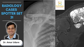
Dr. Ebraheim’s educational animated video describes the condition of osteochondroma. Osteochondroma is a surface lesion that arises from the surface of the bone and continues with the medullary cavity. It arises from a trapped growth plate cartilage that herniates through the cortex and grows via the endochondral ossification beneath the periosteum. The cartilaginous cap produces the bony mass by progressive endochondral ossification. It is the most common benign bone tumor. Presentation: Mass swelling and pain or discomfort. Pain can originate from the bursa or due to mechanical impingement. Pain may also occur due to fracture of the stalk. Locations: Distal femur, proximal tibia, proximal femur, proximal humerus. Osteochondroma can occur however, in any bone in which endochondral ossification occurs. Growth of the osteochondroma parallels the growth of the patient. The lesion will stop growing once the physis closes. X-ray: The cortex of the medullary cavity is continuous. The lesion may be either sessile (uncommon) or pedunculated (common). The base of the lesion is wider with high risk of malignancy in sessile. A pedunculated lesion is a narrow stalk that usually grows away from the joint. Osteochondroma often arises at the site of tendon insertion. The direction of the growth follows along the tendon line. MRI will show the cartilage cap better than an x-ray. The cap is usually 2-3 mm in thickness. It may be 1 cm thick in a grown child. The thicker cap indicates growth, however, it is not a reliable indication of malignant transformation. The patient may have a bursa on top of the lesion and there may be calcified or ossified loose bodies. The cartilaginous cap is made of hyaline cartilage and it has chondrocytes that are benign with a single nucleus usually arranged in clusters and parallel similar to the physis. If the cartilage cap becomes thicker in adults (more than 2 cm), the rule out chondrosarcoma. Multiple hereditary exostoses is a disorder that is characterized by multiple osteochondromas. The lesions are similar in x-ray appearance and histologically to solitary osteochondroma. It may have metaphyseal widening and sessile lesions. It is autosomal dominant with incomplete penetrance in females. It is more common in males. It occurs due to a gene mutation EXT1, EXT2, EXT3. In EXT1, there will be more severe presentation such as more limb malalignment and decreased range of motion of the knee and elbow. EXT1 will have more extostoses and more malignancy than in EXT2. Malignant transformation solitary is less than 1% and multiple is about 10%. Proximal lesions tend to undergo more malignant transformation than distal lesions. If it transforms to malignant, then the lesion will be low grade chondrosarcoma. This usually occurs in the pelvis. Presentation: Short stature (shortened femur), skeletal deformities (site of limb deformities is usually the knee, forearm and ankle), coxa valga, knee valgus with short fibula and patellar dislocation, and ankle valgus due to a shortened fibula. In the upper extremity, there may be radial bowing with the ulna shortened, the radial head dislocated and ulnar deviation of the hand (treated by an osteotomy and exostosis excision). The condition is similar to Madelung’s deformity. Malignant transformation: Clinically, any new pain or sudden increase in size of the lesion will signal low grade chondrosarcoma that usually occurs in the scapula or pelvis. Asses the cartilage cap (MRI will show the cap as a bright sign). It occurs more in the sessile type showing an area of lucency or destruction of the base of the oteochondroma or destruction of the adjacent bone. May also find presence of a calcified soft tissue mass. Usually occurs in older patients. Differential diagnosis includes periosteal osteosarcoma and myositis ossificans. Treatment: Observation if the patient is asymptomatic. Excision after skeletal maturity is usually done if there is a presence of pain, cosmetic deformity or loss of range of motion. Need to be aware of any loss of pronation and supination in the forearm. A wide surgical excision will be done in case of a secondary chondrosarcoma. Become a friend on facebook: / drebraheim Follow me on twitter: https://twitter.com/#!/DrEbraheim_UTMC Donate to the University of Toledo Foundation Department of Orthopaedic Surgery Endowed Chair Fund: https://www.utfoundation.org/foundati...
Comments