Скачать с ютуб Cross sectional and imaging anatomy of the thorax в хорошем качестве
Cross sectional and imaging anatomy of the thorax
10 лет назад
Скачать бесплатно Cross sectional and imaging anatomy of the thorax в качестве 4к (2к / 1080p)
У нас вы можете посмотреть бесплатно Cross sectional and imaging anatomy of the thorax или скачать в максимальном доступном качестве, которое было загружено на ютуб. Для скачивания выберите вариант из формы ниже:
Загрузить музыку / рингтон Cross sectional and imaging anatomy of the thorax в формате MP3:
Если кнопки скачивания не
загрузились
НАЖМИТЕ ЗДЕСЬ или обновите страницу
Если возникают проблемы со скачиванием, пожалуйста напишите в поддержку по адресу внизу
страницы.
Спасибо за использование сервиса savevideohd.ru
Cross sectional and imaging anatomy of the thorax
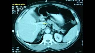
This video deals with the anatomy of the thorax in transverse anatomical and axial CT sections. The video begins with a transverse anatomical section at the root of the neck and continues down to the level of the T10 vertebra where the esophagus passes through the diaphragm. The anatomical sections are arranged to match CT & MRI sections to provide a better understanding of the imaging anatomy of the thorax. The arrangement of thoracic structures is followed in (16) serial transverse sections of the thorax and compared with (10) representative axial CT sections at different levels. 00:00 Introduction 00:30 Section 1 02:47 Section 2 06:11 Section 3 10:37 Axial CT 1 12:02 Section 4 13:43 Axial CT 2 14:20 Section 5 16:11 Axial CT 3 16:54 Section 6, level of T4 vertebra 19:46 Axial CT 4 20:11 Section 7 22:50 Axial CT 5 23:40 Section 8 26:25 Axial CT 6 27:03 Section 9 28:36 Axial CT 7 29:34 Section 10 31:03 Section 11 31:45 Axial CT 8 33:22 Section 12 34:43 Section 13 35:55 Section 14 38:15 Section 15 39:09 Axial CT 9 29:40 Section 16 40:55 Axial CT 10 The anatomical sections are selected from the Visible Human Project. For more information about this project refer to: http://www.nlm.nih.gov/research/visib... A plastic model is used where necessary to explain the 3-D relations necessary to understand the 2-D sections. Presented and edited by Akram Jaffar, Ph.D. This video and its channel are supported by the "Human Anatomy Education" page on Facebook / anatomyeducation After completion of this video session, it is expected that you will be able to identify the anatomical features expected to be seen at vertebral levels T2, 4, 8, and 10. The following structures are identified and followed in transverse anatomical sections or CT axial sections: Vessels: Ascending aorta, descending aorta, arch of aorta, brachiocephalic trunk, left common carotid artery, left subclavian artery, right coronary artery, internal thoracic (mammary) artery, superior vena cava (SVC), inferior vena cava (IVC), azygos vein, right and left brachiocephalic trunks, internal jugular vein, subclavian vein, coronary sinus, pulmonary trunk, right and left pulmonary artery, pulmonary vein. Viscera: Esophagus, lungs, liver, spleen, thymus, stomach, heart: right ventricle, left ventricle, right atrium, right auricle, left atrium, vestibule, infundibulum, apex, interventricular septum, trabeculae carneae, papillary muscle, moderator band, right atrioventricular orifice, trachea, right and left main bronchus, right upper lobe bronchus, hilar lymph node, carinal lymph nodes, right and left domes of the diaphragm. Musculoskeletal structure: Ribs, costal cartilages, sternum, scapula, clavicle, sternoclavicular joint, intercostal muscles, serratus anterior, pectoralis major, pectoralis minor, vertebral body, transverse process, spinous process, vertebral canal, spinal cord, costovertebral joint, costotransverse joint, intervertebral disc. Related videos: Relations at the superior mediastinum, simplified sketches • Relations at the superior mediastinum... Cross-sectional and imaging anatomy of the abdomen • Cross sectional and imaging anatomy o... Cross-sectional anatomy of the female pelvis and perineum • Cross sectional anatomy of the female... Related accounts Twitter / akramjaffar Facebook / anatomyeducation SlideShare http://www.slideshare.net/AkramJaffar LinkedIn / akram-abo. . Research gate https://www.researchgate.net/profile/... Medtube https://medtube.net/users/akram-jaffar Instagram / akramjaffar Academia https://dal.academia.edu/AkramJaffar
Comments