Скачать с ютуб Achilles Tendon: how to Ultrasound Scan в хорошем качестве
Achilles Tendon: how to Ultrasound Scan
6 лет назад
Из-за периодической блокировки нашего сайта РКН сервисами, просим воспользоваться резервным адресом:
Загрузить через ClipSave.ruСкачать бесплатно Achilles Tendon: how to Ultrasound Scan в качестве 4к (2к / 1080p)
У нас вы можете посмотреть бесплатно Achilles Tendon: how to Ultrasound Scan или скачать в максимальном доступном качестве, которое было загружено на ютуб. Для скачивания выберите вариант из формы ниже:
Загрузить музыку / рингтон Achilles Tendon: how to Ultrasound Scan в формате MP3:
Если кнопки скачивания не
загрузились
НАЖМИТЕ ЗДЕСЬ или обновите страницу
Если возникают проблемы со скачиванием, пожалуйста напишите в поддержку по адресу внизу
страницы.
Спасибо за использование сервиса savevideohd.ru
Achilles Tendon: how to Ultrasound Scan
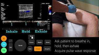
How to scan the Achilles Tendon using Ultrasound. In this video, sonographer Milton Delves, demonstrates the achilles tendon ultrasound technique. Probe positioning: Make sure there is enough gel right over the heels especially when the skin surface is not flat to avoid any air gaps between the probe and the skin surface. Place the probe longitudinal to show a long axis view of the Achilles tendon then slide up toward the calf muscles and then down along the axis towards the insertion into the heel bone. By moving the feet up and down you can evaluate the tendon while in motion. The Achilles tendon is a cylinder like structure and should always appear hyperechoic with homogeneous fibers pattern along its axis. Angle the probe 90 degrees to get the transverse (Short) view and repeat the scan from the beginning of the calf muscles to the insertion over the calcaneus to make sure no findings were missed in the longitudinal view. Any swelling will be identified by an increase in tendon thickness accompanied by a mild hypoechoic (dark) areas and slight loss of fibrillar pattern. Also pay attention to any areas of fluid collection which may occur around the tendon or in the bursa. Finally the power or colour doppler should be used to check for areas of hyperaemia.
Comments