Скачать с ютуб Diabetic foot (mechanism of disease) в хорошем качестве
Diabetic foot (mechanism of disease)
1 год назад
Из-за периодической блокировки нашего сайта РКН сервисами, просим воспользоваться резервным адресом:
Загрузить через ClipSave.ruСкачать бесплатно Diabetic foot (mechanism of disease) в качестве 4к (2к / 1080p)
У нас вы можете посмотреть бесплатно Diabetic foot (mechanism of disease) или скачать в максимальном доступном качестве, которое было загружено на ютуб. Для скачивания выберите вариант из формы ниже:
Загрузить музыку / рингтон Diabetic foot (mechanism of disease) в формате MP3:
Если кнопки скачивания не
загрузились
НАЖМИТЕ ЗДЕСЬ или обновите страницу
Если возникают проблемы со скачиванием, пожалуйста напишите в поддержку по адресу внизу
страницы.
Спасибо за использование сервиса savevideohd.ru
Diabetic foot (mechanism of disease)
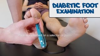
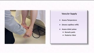
ERRATA (thank you to commenters): Claw toe has dip flexion (not extension). This is a flowchart on the diabetic foot, covering the etiology, pathophysiology, and manifestations. ADDITIONAL TAGS: Risk factors / SDOH Cell / tissue damage Structural factors Medicine / iatrogenic Infectious / microbial Biochem. / metabolic Immunology / inflammation Signs / symptoms Tests / imaging / labs Environmental / exposure Nervous system pathology Flow physiology Pathophysiology Etiology Manifestations Diabetic foot Foot ulcers: skin breakdown with possible surrounding tissue necrosis Diabetic foot ulcer, classified as … Neuropathic ulcers Neuroischemic ulcers Ischemic ulcers Peripheral sensory neuropathy Autonomic neuropathy Peripheral artery disease Microvascular changes Diabetes mellitus Chronic hyperglycemia Sorbitol accumulation in cells Ulcers at sites of repetitive stress/trauma: bony abnormalities, bottom of foot (metatarsal bones or heel) Ulcers on the toes or lateral foot Usually painless +/- sensory loss, motor weakness +/- cool foot, no palpable pulses Infection Trauma Calluses ↑ rates of … Hospitalization Amputation Death Skin and soft tissue infection In ~50% of ulcers Staphylococcus spp. Streptococcus spp. Typically polymicrobial Most common causative pathogens: Edema Induration Erythema 0.5 cm Tenderness Warmth Purulent exudate ↓ cytokine production Defects in phagocytosis Immune cell dysfunction Diabetes mellitus impairs immune system Diabetic foot osteomyelitis Ulcer overlying bony prominence, exposed bone Ulcer size 2 cm2 and / or ulcer depth 3 mm Chronic, treatment-resistant ulcer Positive probe-to-bone test Markedly increased ESR (70 mm/hour) Leukocytosis ↓ intrinsic muscle volume Thickening of plantar aponeurosis Bone destruction Subluxation / dislocation Gas gangrene By Heather Murphy-Lavoie - https://www.ncbi.nlm.nih.gov/books/NB..., CC BY 4.0, https://commons.wikimedia.org/w/index... Hammer toe: PIP joint flexion; +/- DIP joint extension, +/- MTP joint hyperextension Claw toe: PIP joint flexion, DIP joint extension, and MTP joint hyperextension Inflammation: swelling, warmth, erythema Diabetic neuropathic arthropathy (Charcot foot) Mild to moderate pain Midfoot collapse (rocker- bottom foot deformity) Osteolysis → fractures Painless bony deformities
Comments