Скачать с ютуб Picture tests in practical histology of lymphatic tissue part 1 в хорошем качестве
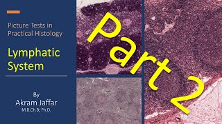
Picture tests in practical histology of lymphatic tissue part 1
8 месяцев назад
Скачать бесплатно Picture tests in practical histology of lymphatic tissue part 1 в качестве 4к (2к / 1080p)
У нас вы можете посмотреть бесплатно Picture tests in practical histology of lymphatic tissue part 1 или скачать в максимальном доступном качестве, которое было загружено на ютуб. Для скачивания выберите вариант из формы ниже:
Загрузить музыку / рингтон Picture tests in practical histology of lymphatic tissue part 1 в формате MP3:
Если кнопки скачивания не
загрузились
НАЖМИТЕ ЗДЕСЬ или обновите страницу
Если возникают проблемы со скачиванием, пожалуйста напишите в поддержку по адресу внизу
страницы.
Спасибо за использование сервиса savevideohd.ru
Picture tests in practical histology of lymphatic tissue part 1
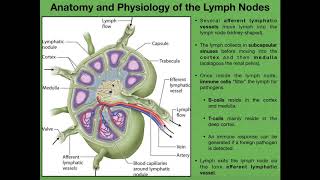
After watching this video, you will be able to: Identify the histological organization of the THYMUS: interlobular septum, thymic (Hassall's) corpuscles LYMPH NODES: outer cortex, inner cortex, medullary cord, medullary sinus, trabecula, afferent lymphatic vessel, efferent lymphatic vessel, hilum, germinal centre, secondary lymphatic nodule. SPLEEN: white pulp, red pulp, central artery mucosal-associated lymphoid tissue (MALT): lingual tonsil and palatine tonsil. Relate structure to function. Video sections 00:36 Q1 What is a primary lymphoid tissue? 03:59 Q2 Where is the main site of removal of worn-out RBCs? 07:16 Q3 Which lymphoid organ has an endocrine function? 10:20 Q4 Which lymphoid organ is considered a filter of blood? Related videos • Histology of the lymphatic system • ANAT1010_27_lymphatic system • Picture tests in practical histology ... Presented and edited by Dr. Akram Jaffar, Ph.D. This video and its channel are supported by the "Human Anatomy Education" Page on Facebook / anatomyeducation Related accounts Twitter / akramjaffar Facebook / anatomyeducation SlideShare http://www.slideshare.net/AkramJaffar LinkedIn / akram-abo. . Research gate https://www.researchgate.net/profile/... Medtube https://medtube.net/users/akram-jaffar Instagram / akramjaffar Academia https://dal.academia.edu/AkramJaffar
Comments