Скачать с ютуб Case of the Week: Amebic Liver Abscess (Ultrasound & CT) в хорошем качестве
Case of the Week: Amebic Liver Abscess (Ultrasound & CT)
2 года назад
Скачать бесплатно Case of the Week: Amebic Liver Abscess (Ultrasound & CT) в качестве 4к (2к / 1080p)
У нас вы можете посмотреть бесплатно Case of the Week: Amebic Liver Abscess (Ultrasound & CT) или скачать в максимальном доступном качестве, которое было загружено на ютуб. Для скачивания выберите вариант из формы ниже:
Загрузить музыку / рингтон Case of the Week: Amebic Liver Abscess (Ultrasound & CT) в формате MP3:
Если кнопки скачивания не
загрузились
НАЖМИТЕ ЗДЕСЬ или обновите страницу
Если возникают проблемы со скачиванием, пожалуйста напишите в поддержку по адресу внизу
страницы.
Спасибо за использование сервиса savevideohd.ru
Case of the Week: Amebic Liver Abscess (Ultrasound & CT)
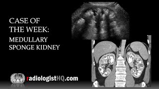
In this radiology lecture, we discuss the ultrasound and CT appearance of amebic liver abscess. Key points include: 1) Caused by Entamoeba histolytica infection. 2) Endemic in Africa, Southeast Asia, and Central & South America. 3) More common in males. 4) Presents as right upper quadrant pain, fever and hepatomegaly. 5) Both amebic and pyogenic (bacterial) abscesses can have a layered wall with the “double target” or “double rim” sign. 6) Amebic more likely to be unilocular (septations present in 30%) without “cluster” sign typical of multiloculated pyogenic abscess. 7) Amebic more likely solitary, pyogenic more likely multiple. 8) Can be treated medically (metronidazole), but if diagnosis uncertain, if there is failed response to medical therapy, or if large abscess at risk for rupture = aspiration. Bächler P, Baladron MJ, Menias C, et al. Multimodality Imaging of Liver Infections: Differential Diagnosis and Potential Pitfalls. RadioGraphics 2016 36:4, 1001-1023. Click the Community tab or follow on social media for bonus teaching material posted throughout the week! Website: http://www.radiologistHQ.com Video Podcast: http://bit.ly/radiologistHQ Instagram: / radiologisthq Facebook: / radiologistheadquarters Twitter: / radiologisthq
Comments