Скачать с ютуб Histology of Uterus-Proliferative Phase or Follicular Phase в хорошем качестве
Histology of Uterus-Proliferative Phase or Follicular Phase
3 года назад
Скачать бесплатно Histology of Uterus-Proliferative Phase or Follicular Phase в качестве 4к (2к / 1080p)
У нас вы можете посмотреть бесплатно Histology of Uterus-Proliferative Phase or Follicular Phase или скачать в максимальном доступном качестве, которое было загружено на ютуб. Для скачивания выберите вариант из формы ниже:
Загрузить музыку / рингтон Histology of Uterus-Proliferative Phase or Follicular Phase в формате MP3:
Если кнопки скачивания не
загрузились
НАЖМИТЕ ЗДЕСЬ или обновите страницу
Если возникают проблемы со скачиванием, пожалуйста напишите в поддержку по адресу внизу
страницы.
Спасибо за использование сервиса savevideohd.ru
Histology of Uterus-Proliferative Phase or Follicular Phase
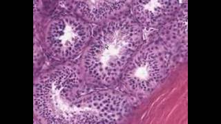
Proliferative Phase Histology. Estrogenic stimulation causes the endometrium to regenerate and proliferate. In the early proliferative phase the glands are straight and narrow and the glandular epithelium is cubo-columnar. Nuclear chromatin appears dispersed and mitotic figures are present. The stromal cells also show mitotic activity and have ill-defined borders . In the late proliferative phase the glands increase in size and appear tortuous with pseudostratification of the epithelium showing nuclei at different levels. The stromal cells are small and spindle-shaped similar to predecidual cells. Cytology. In the early proliferative phase, EBT show glandular cells in cohesive monolayered sheets. They sometimes appear as straight or twisted tubular structures resembling glove fingers irregularly sheared at the ends, the so-called glove-finger pattern. Almost all the tubular fragments are open at both ends (open type), but a few are closed at one end (closed type) and have a cup-like, a half moon, or spherical shape . The nuclei are of uniform size and shape and have granular chromatin and distinct micronucleoli. Loose aggregates of stromal cells, which have oval nuclei and poorly defined cytoplasm, can be identified. In the late proliferative phase, sheets of endometrial cells are highly cellular with nuclear crowding, denser nuclear chromatin and frequent mitotic figures. Tubular structures are small and a glove-finger pattern is frequently seen The uterine corpus is composed of a modified mucosa known as the endometrium, a fibromuscular wall called the myometrium, and a serosal lining. The uterine mucosa can be divided into two regions: the mucosa of the lower uterine segment (LUS) (isthmus) and the mucosa of the corpus proper The mucosa of the LUS, located between the endocervix and endometrium, is thinner than that of the fundus and its glands respond only slightly to hormonal stimulation. There is a gradual morphologic transition from the isthmic mucosa to the endocervical mucosa. During the reproductive years the endometrium of the corpus proper undergoes regular cyclic changes as a response to the release of the ovarian hormones, estrogen and progesterone. The endometrium consists of simple tubular glands set in a cellular vascular stroma. It is composed of a thin basal layer The structure and activity of a functional endometrium reflect the pattern of ovarian hormone secretion. The histologic types of glandular cells are columnar or cuboid. The endometrium undergoes regular growth and maturation and when the cycle ends, in the absence of pregnancy, shedding occurs followed by regeneration. The average duration of the cycle is 28 days. In a normal cycle the postovulatory phase lasts 14 days. Changes in the length of the cycle are usually due to the duration of the proliferative phase, which can vary from 8 to 21 days
Comments