Скачать с ютуб How To Scan The Uterine Artery | Doppler Ultrasound Probe Positioning | Transducer Placement USG в хорошем качестве
How To Scan The Uterine Artery | Doppler Ultrasound Probe Positioning | Transducer Placement USG
10 месяцев назад
Скачать бесплатно How To Scan The Uterine Artery | Doppler Ultrasound Probe Positioning | Transducer Placement USG в качестве 4к (2к / 1080p)
У нас вы можете посмотреть бесплатно How To Scan The Uterine Artery | Doppler Ultrasound Probe Positioning | Transducer Placement USG или скачать в максимальном доступном качестве, которое было загружено на ютуб. Для скачивания выберите вариант из формы ниже:
Загрузить музыку / рингтон How To Scan The Uterine Artery | Doppler Ultrasound Probe Positioning | Transducer Placement USG в формате MP3:
Если кнопки скачивания не
загрузились
НАЖМИТЕ ЗДЕСЬ или обновите страницу
Если возникают проблемы со скачиванием, пожалуйста напишите в поддержку по адресу внизу
страницы.
Спасибо за использование сервиса savevideohd.ru
How To Scan The Uterine Artery | Doppler Ultrasound Probe Positioning | Transducer Placement USG
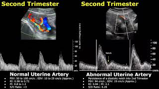
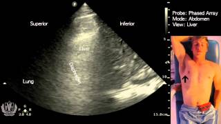
How To Scan The Uterine Artery | Doppler Ultrasound Probe Positioning | Transducer Placement USG - Patient Positioning: The patient should be lying in the supine position - Probe Selection: Choose a curved array transducer, commonly referred to as a "convex" or "abdominal" probe. A frequency of 2-5 MHz is typically used for transabdominal imaging. - Probe Placement: Place the transducer on the lower abdomen, just above the pubic symphysis, with the probe marker oriented towards the patient's head. Sweep the probe side-to-side and up-and-down to visualize the uterus in both the sagittal and transverse planes. - Identifying Uterine Arteries: The uterine arteries are usually identified as they cross over the external iliac arteries. They can be seen as pulsatile vessels on color Doppler, at the internal cervical os, the junction between the uterine body and cervix. The uterine arteries are located on either side of the uterus, so visualizing the uterus itself can help identify the position of these vessels. The uterine arteries run along the cervix, so identifying the cervix can be a useful landmark. Color Doppler can be used to distinguish the uterine arteries from surrounding structures. The uterine arteries show pulsatile blood flow, which can help differentiate them from nearby veins. - Doppler Imaging: To evaluate blood flow, switch to Doppler mode. Place the Doppler sample volume over the uterine artery, and adjust the scale to capture the pulsatile flow. Measure peak systolic and end-diastolic velocities, as well as the resistance index (RI) and pulsatility index (PI) if required. - Sampling Gate: 2mm - Angle Of Insonation: Less than 30 degrees - 3 consecutive waveforms are sufficient for spectral doppler evaluation #uterinearterydoppler #uterineartery #uterinearteryultrasound
Comments