Скачать с ютуб Osteology of Head & Neck - Temporal Bone в хорошем качестве
Osteology of Head & Neck - Temporal Bone
5 лет назад
Скачать бесплатно Osteology of Head & Neck - Temporal Bone в качестве 4к (2к / 1080p)
У нас вы можете посмотреть бесплатно Osteology of Head & Neck - Temporal Bone или скачать в максимальном доступном качестве, которое было загружено на ютуб. Для скачивания выберите вариант из формы ниже:
Загрузить музыку / рингтон Osteology of Head & Neck - Temporal Bone в формате MP3:
Если кнопки скачивания не
загрузились
НАЖМИТЕ ЗДЕСЬ или обновите страницу
Если возникают проблемы со скачиванием, пожалуйста напишите в поддержку по адресу внизу
страницы.
Спасибо за использование сервиса savevideohd.ru
Osteology of Head & Neck - Temporal Bone
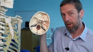
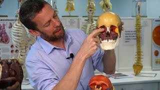
Temporal bone - a pair or irregular pneumatic bones. The parts - Squamous Petro-mastoid Tympanic Styloid process Squamous part - presents External surface Internal surface Superior border Antero inferior border External surface forms floor of temporal fossa gives attachment to temporlis muscle extends 1.5 cms below the supramastoid crest External surface shows zygomatic process with anterior and posterior roots. It unites with temporal process of zygomatic process completing zygomatic arch Upper border of zygomatic arch gives attachment to galea aponeurotica, temporal fascia Lower border and inner surface gives attachment to masseter muscle Internal Surface or cerebral surface shows impressions of gyri of temporal lobes of cerebrum, impressions of middle temporal vessels Superior border articulates with middle 1/3 of lower border parietal bone Anteroinferior border articulates with squamous part of greater wing of sphenoid Mastoid part - presents External surface, inner surface superior & posterior border and a mastoid process External Surface give attachment to Sternocleidomastoid muscle, splenius capitis, longissimus capitis muscles from before backwards It presents mastoid foramen which transmits emmissary vein connecting the sigmoid sinus with the posterior auricular vein. Internal surface presents sulcus for sigmoid sinus superior border articulates with posterior 1/3 of lower border of parietal bone Posterior border articulates with lateral border of occipital bone Tympanic Part presents anterior & Posterior surfaces Superior inferior & lateral borders Anterior surface forms the non articular part of mandibular fossa Posterior surface forms anterior wall , floor & lower part of external acoustic meatus Superior border meets the squamous part at the squamotympanic fissure This fissure is divided by the downturned edge of tegmen tympani into petrotympanic fissure and petro squamous fissure Structures passing through the petro tympaic fissure - Chorda tympani nerve, anterior tympanic artery, anterior ligament of malleus Inferior border splits to enclose the styloid process (vaginal process) Lateral border forms the bony ring where cartilage of ear are attached Petrous part Presents anterior, posterior & inferior surfaces anterior, superior & posterior borders An apex & a base The apex is irregular, presents an impressions for trigeminal ganglion, covered by meckel's cave. It also shows the anterior opening of carotid canal. It forms the posterolateral boundary for the foramen lacerum The base blends with squamous & mastoid part Anterior surface presents arcuate fossa, tegmen tympani Posterior surface presents internal acoustic meatus, oblique slit - vestibule of aqueduct, subarcuate fossa Inferior surface presents attachments to levator veli palatini , lateral to this forms sulcus tubae along with greater wing of sphenoid which lodges the cartilaginous part of auditory tube Lateral to this is the jugular fossa which lodges the supeior bulb of internal jugular vein infront to this fossa presents impression of inferior ganglion of glossopharyngeal nerve Styloid process - downward projection of 2.5 cms long - develop from the dorsal part pharyngeal arch It gives attachment to 3 muscles - styloglossus, stylopharyngeus, stylohyoid muscles 2 ligaments - stylohoid & stylomandibular ligament Related laterally to parotid gland with external carotid artery & facial nerve medially related to internal jugular vein & internal carotid artery with last four cranial nerves inbetween them Follow me in blogspot - https://human-anatomylessons.blogspot... _________________________________________________________________________________________________________________ Visit my blogs - https://humananatomyonline.in/ Contact me @ https://t.me/humananatomylessons
Comments