Скачать с ютуб Esophageal Cancer Diagnosis and Staging в хорошем качестве
Esophageal Cancer Diagnosis and Staging
3 года назад
Скачать бесплатно Esophageal Cancer Diagnosis and Staging в качестве 4к (2к / 1080p)
У нас вы можете посмотреть бесплатно Esophageal Cancer Diagnosis and Staging или скачать в максимальном доступном качестве, которое было загружено на ютуб. Для скачивания выберите вариант из формы ниже:
Загрузить музыку / рингтон Esophageal Cancer Diagnosis and Staging в формате MP3:
Если кнопки скачивания не
загрузились
НАЖМИТЕ ЗДЕСЬ или обновите страницу
Если возникают проблемы со скачиванием, пожалуйста напишите в поддержку по адресу внизу
страницы.
Спасибо за использование сервиса savevideohd.ru
Esophageal Cancer Diagnosis and Staging
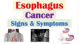
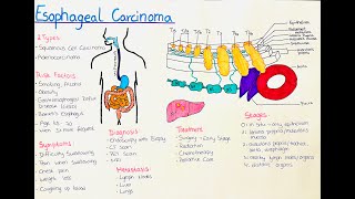
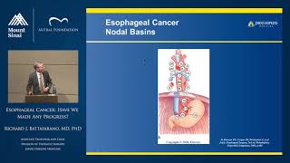
This video will help you understand the diagnosis and staging of esophageal cancer. In this video you’ll learn about: • Definitions of cancer Terms • Staging of esophageal cancer • Diagnostic Tests The esophagus is a hollow muscular tube which connects the throat to the stomach. For many patients, the first symptom they have is difficulty swallowing or pain in swallowing. The difficulty swallowing can be due to a lump, also known as a tumor Cancer Terms for Esophageal Cancer A tumor is an abnormal growth Tumors can be either benign or malignant • A benign tumor of the esophagus may grow over time and cause obstruction and make it difficult to swallow, but it won’t ever spread anywhere else Malignant Tumors of the Esophagus = Cancer • A malignant tumor of the esophagus can cause obstruction • A malignant tumor also has the potential to spread elsewhere in the body. • Esophageal cancer is another term for a malignant tumor of the esophagus • We will use the term cancer and tumor interchangeably in this video In most cases, the diagnosis of esophageal cancer is made by endoscopy, also called an EGD. Under sedation, a flexible scope is passed through the mouth into the esophagus, which allows viewing the inside of the esophagus. Staging If the biopsy shows cancer, the next step is staging. Staging is the process of finding out the size of the tumor and whether or not there has been spread to the lymph nodes or other places in the body. Once the stage has been determined, it will be possible to determine the best therapy The wall of the esophagus has multiple layers. Surrounding the esophagus are lymph nodes. The purpose of lymph nodes is to filter the blood and help fight infections, but in some cases cancers in the esophagus can spread to the lymph nodes In its earliest stages, cancer of the esophagus starts on the inner, or most superficial layer, called with mucosa. With time, however, esophageal cancer can continue to grow and invade deeper into the wall of the esophagus The deeper the cancer invades into the wall of the esophagus, the more likely it is that cancer cells can spread to the lymph nodes If cancer cells spread to the lymph nodes, there is a chance that some cells will break off and spread to the liver or lungs. Metastasis is spread to other parts of the body such as liver, lungs, or bone. The stage consists of 3 parts: T for Tumor (How deep has the cancer invaded into the wall of the esophagus?) N for Nodes (Has the cancer spread to the lymph nodes? How many?) M for Metastasis (Has the cancer spread to other parts of the body?) T1 tumor involves the top layers of the esophagus T1a tumor involves the mucosa T1b tumor involves the submucosa T2 tumor invades into the muscular layer T3 tumor invades all the way through the muscular layer T4 tumor invades into nearby structures such as the aorta or the airway As a general rule, if someone with esophageal cancer has difficulty swallowing, the tumor is usually a T3 The N classification refers to the lymph nodes N0 – No lymph nodes N1 – 1 or 2 lymph nodes involved N2 – 3-6 nodes involved N3 – 7 or more nodes involved The M classification refers to metastasis Metastasis = spread of cancer to other organs such as the lungs, liver, or bone M0 – No signs of spread to other organs M1 – Spread to other organs Diagnostic Tests When you meet with your doctors, one the first things they will do is to come up with a plan for testing that is tailored to you and your particular tumor. A CT scan is usually the first test for staging. This will show whether there is any signs of metastasis or spread to other organs such as the lung, liver, or bone A PET scan is a specialized scan which combines a CT scan with an injection of a small amount of tracer which lights up areas of cancer. In cases where it is important to know about the exact size of the tumor, an endoscopic ultrasound exam (EUS) can be done. This procedure is similar to an EGD, but the endoscope has an ultrasound sensor on the end of the scope which produces an image of the tumor In some cases, particularly for cancers in the stomach, it is important to look for signs of spread in the abdominal cavity. In some situations, cancers can spread in the abdominal cavity but the areas are so small they don’t show up on a CT scan. In these cases, a laparoscopy is helpful. Laparoscopy is a surgical procedure done under a general anesthetic. Several incisions ¼” long are made, and a telescope is inserted into the abdominal cavity. This allows an examination of the abdominal cavity to look for signs of spread. The procedure is usually done as an outpatient, so you can go home the same day.
Comments