Скачать с ютуб Langerhans Histiocytosis, Pemphigus, Tattoo, Tick, Mycosis Fungoides, &Keloid under the microscope. в хорошем качестве
Langerhans Histiocytosis, Pemphigus, Tattoo, Tick, Mycosis Fungoides, &Keloid under the microscope.
3 года назад
Из-за периодической блокировки нашего сайта РКН сервисами, просим воспользоваться резервным адресом:
Загрузить через ClipSave.ruСкачать бесплатно Langerhans Histiocytosis, Pemphigus, Tattoo, Tick, Mycosis Fungoides, &Keloid under the microscope. в качестве 4к (2к / 1080p)
У нас вы можете посмотреть бесплатно Langerhans Histiocytosis, Pemphigus, Tattoo, Tick, Mycosis Fungoides, &Keloid under the microscope. или скачать в максимальном доступном качестве, которое было загружено на ютуб. Для скачивания выберите вариант из формы ниже:
Загрузить музыку / рингтон Langerhans Histiocytosis, Pemphigus, Tattoo, Tick, Mycosis Fungoides, &Keloid under the microscope. в формате MP3:
Если кнопки скачивания не
загрузились
НАЖМИТЕ ЗДЕСЬ или обновите страницу
Если возникают проблемы со скачиванием, пожалуйста напишите в поддержку по адресу внизу
страницы.
Спасибо за использование сервиса savevideohd.ru
Langerhans Histiocytosis, Pemphigus, Tattoo, Tick, Mycosis Fungoides, &Keloid under the microscope.
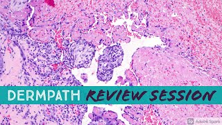
Recording of random dermpath case review session with my dermatology residents and dermpath fellows on 7/16/2021. Thanks to my friend Dr. Tammie Ferringer for letting me use these great slides from her collection! Sorry, the slide viewer system is not able to share the digital slides publicly. Topics Discussed (click timestamp to go to that part of video): Keloid with injected steroid material (Triamcinolone) 0:00 (plus bonus findings of actinic keratosis and epidermodysplasia verruciformis +/- SCC in situ) Suture 6:04 Currettings at least actinic keratosis vs SCC 9:16 Nail psoriasis 15:47 Tattoo reaction 18:15 Porokeratosis ptychotropica 25:30 Talon noir 28:52 Condyloma 30:12 with cautery artifact 34:55 Nevus of Ota (dermal melanocytosis) 37:00 Curling iron burn (freeze injury would show same pattern) 39:43 Tick 43:16 Langerhans cell histiocytosis (vs mastocytosis) 47:15 Supernumerary/accessory nipple 51:00 Pemphigus vulgaris 52:23 Tumoral melanosis (completely regressed melanoma or nevus) 55:06 Pink Over Blue Trick (New Collagen Replacing Solar Elastosis as Clue for Regression) 57:50 BCC that fell out of tissue during processing artifact 1:00:00 Sebaceous carcinoma of the eyelid with Adipophilin 1:01:51 Sarcoid 1:06:22 Radiation dermatitis 1:09:52 Cellular blue nevus 1:12:39 Tissue reaction to ferruginous iron-containing foreign body (positive Wolverine Sign!) 1:15:02 Pagetoid reticulosis variant of mycosis fungoides/cutaneous T-cell lymphoma (Woringer-Kolopp disease) 1:16:37 Normal Eyelid Histology (plus a nevus) 1:18:16 Gout 1:19:08 Silicone granuloma 1:19:40 Gel foam 1:20:55 Other useful videos and links: EDV video: https://kikoxp.com/posts/4425 Mycosis Fungoides basics video: https://kikoxp.com/posts/4131 Squamous cell carcinoma & actinic keratosis 101: https://kikoxp.com/posts/3771 Porokeratosis video: https://kikoxp.com/posts/4376 Verruca vulgaris 101: https://kikoxp.com/posts/6126 Tungiasis: Video https://kikoxp.com/posts/2341 & digital slide: https://kikoxp.com/posts/2338 Sebaceous Lesions 101 video: https://kikoxp.com/posts/6344 Cellular Blue Nevus vs Deep Penetrating Nevus video: https://kikoxp.com/posts/3917 Gout video: https://kikoxp.com/posts/3799 A complete organized library of all my videos, digital slides, pics, & sample pathology reports is available here: https://kikoxp.com/posts/5084 (dermpath) & https://kikoxp.com/posts/5083 (bone/soft tissue sarcoma pathology). Please check out my Soft Tissue Pathology & Dermatopathology survival guide textbooks: http://bit.ly/2Te2haB This video is geared towards medical students, pathology or dermatology residents, or practicing pathologists or dermatologists. Of course, this video is for educational purposes only and is not formal medical advice or consultation. Presented by Jerad M. Gardner, MD. Please subscribe to my channel to be notified of new pathology teaching videos. Follow me on: Snapchat: JMGardnerMD Twitter: @JMGardnerMD Instagram: @JMGardnerMD Kiko: https://kikoxp.com/profile/jerad_gard... Facebook: / jmgardnermd
Comments