Скачать с ютуб Acute Cholecystitis (2/3) в хорошем качестве
Acute Cholecystitis (2/3)
4 года назад
Из-за периодической блокировки нашего сайта РКН сервисами, просим воспользоваться резервным адресом:
Загрузить через ClipSave.ruСкачать бесплатно Acute Cholecystitis (2/3) в качестве 4к (2к / 1080p)
У нас вы можете посмотреть бесплатно Acute Cholecystitis (2/3) или скачать в максимальном доступном качестве, которое было загружено на ютуб. Для скачивания выберите вариант из формы ниже:
Загрузить музыку / рингтон Acute Cholecystitis (2/3) в формате MP3:
Если кнопки скачивания не
загрузились
НАЖМИТЕ ЗДЕСЬ или обновите страницу
Если возникают проблемы со скачиванием, пожалуйста напишите в поддержку по адресу внизу
страницы.
Спасибо за использование сервиса savevideohd.ru
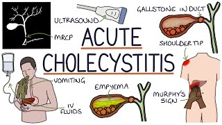
Acute Cholecystitis (2/3)
0:00 Anatomy 1:57 Pathophysiology 7:23 Complications 12:42 Symptoms, Physical Exam Findings, Labs 17:21 RUQ Ultrasound 23:08 HIDA Scan 32:44 Treatment Handwritten lectures about Cholecystitis for USMLE Step 1 and USMLE Step 2 ANATOMY OF BILIARY TRACT IN PATIENTS WITH CHOLECYSTITIS The gallbladder is connected to the cystic duct. The cystic duct combines with a hepatic duct which forms the common bile duct. It combines with the pancreatic duct to excrete through the sphincter of Oddi. Generally, Cholecystitis occurs when a stone is lodged into the cystic duct PATHOLOGY OF CHOLECYSTITIS CALCULOUS CHOLECYSITIS In patients with cholecystitis, a gallstone is formed in the gall bladder which obstructs the cystic duct. As the gallbladder is contracting, the gallbladder becomes inflamed and this is called cholecystitis secondary to a stone. Edema is formed around the gallbladder and the release of prostaglandins increases contraction and this creates the typical pain of cholecystitis. Afterward, a secondary infection can occur due to E. Coli, Enterococcus, Enterobacter, and Klebsiella. The gallstone can lodge into the common bile duct. This is no longer called cholecystitis, rather it is known as choledocholithiasis. ACALCULOUS CHOLECYSTITIS In acalculous cholecystitis, there is inflammation of the gallbladder without the presence of the stone. Usually, there is stasis or ischemia secondary to some severe diseases such as cardiac arrest, sepsis, or burns. However, cholecystitis can also occur due to a primary infection of Ascaris, echinococcus, Brucella, Coxiella, cryptosporidium, isosprora, TB, vibrio, salmonella, Hepatitis A, and Hepatitis B Virus. Immunosuppressed patients, such as AIDS and leukemia can also lead to acalculous cholecystitis. COMPLICATION OF CHOLECYSTITIS The most common complication is gangrenous cholecystitis which occurs in 20 percent of cases. The most common risk factors is elderly, diabetes, delaying seeking treatment. Gangrenous cholecystitis is a life-threatening emergency and requires immediate surgery. PERFORATION After gangrenous cholecystitis patients can have a perforation. This may be associated with a pericholecystic abscess which is palpable. Free perforation will cause generalized peritonitis and associated with high mortality. EMPHYSEMATOUS CHOLECYSTITIS This is when gas-forming bacteria infect the gallbladder wall. Common organisms are staph, strep, Pseudomonas, and klebsiella. This form of cholecystitis is very dangerous. Often confused for overlying bowel gas. BOWEL OBSTRUCTION A fistula can form between the gallbladder and interesting known as cholecystoenteric fistula. A stone greater than 2.5cm can lodge into the ileocecal vale and cause an obstruction. SYMPTOMS OF CHOLECYSTITIS Pain is typically in RUQ radiating to the back or right shoulder. Pain is persistent an lasts 4 to 6 hours. Associated with eating fatty meals and movements. Also will have fever, nausea, vomiting. On examination, the patient will have peritoneal signs and will not try to move. There may also be involuntary or voluntary guarding. Murphy's sign is specific for cholecystitis as well. On labs, the patient will have an elevated white blood count. Also check ALP, bilirubin, AST, ALT to rule out the involvement of the common bile duct and liver. In the RUQ ultrasound, there are two views, longitudinal and transverse views to assess for cholecystitis. Typically you will see an enlargement of the gallbladder wall greater than 3mm with pericholecystic fluid. However, for cholecystitis, the sonographic murphy's sign and visualization of the gallstone is more specific. It is important to look for common bile duct size as if greater then 6mm it is suggestive of choledocholithiasis rather than cholecystitis. If there is suspicion for emphysematous cholecystitis, you will possibly see air in the gallbladder wall. HIDA SCAN If ultrasound is unequivocal for cholecystitis a HIDA Scan also known as Hepatic Iminodiacetic Acid or Cholescintigraphy. In this scan technetium-99 is injected and taken up by the liver. Eventually, it drains into the bile duct. If the cystic duct is patent the gallbladder will show, which rules out cholecystitis. If you don't see the gallbladder than rules out cholecystitis. However, you should wait 3-4 hours or administer morphine augmentation before diagnosing cholecystitis. TREATMENT Start with supportive care with hydration, NPO, Analgesia with NSAIDs, and Opioids. Anti-biotics typically given are Flagyl and ceftazidime, and fluoroquinolone. If the patient is a high risk, then you can go broader with zosyn or ertapenem. Consider vanco if concern for hospital-acquired cholecystitis. Emergency cholecystectomy is performed if cholecystitis is complicated or there is disease progression in uncomplicated cholecystitis. If uncomplicated perform surgery within 3 days. If high risk considers drainage.
Comments


![Gallbladder: Cholelithiasis vs Cholecystitis vs Choledocholithiasis vs Cholangitis [Made Easy]](https://i.ytimg.com/vi/Ccs7DAbzvpE/mqdefault.jpg)

