Скачать с ютуб Neuroanatomy - The spinal cord в хорошем качестве
Neuroanatomy - The spinal cord
9 лет назад
Скачать бесплатно Neuroanatomy - The spinal cord в качестве 4к (2к / 1080p)
У нас вы можете посмотреть бесплатно Neuroanatomy - The spinal cord или скачать в максимальном доступном качестве, которое было загружено на ютуб. Для скачивания выберите вариант из формы ниже:
Загрузить музыку / рингтон Neuroanatomy - The spinal cord в формате MP3:
Если кнопки скачивания не
загрузились
НАЖМИТЕ ЗДЕСЬ или обновите страницу
Если возникают проблемы со скачиванием, пожалуйста напишите в поддержку по адресу внизу
страницы.
Спасибо за использование сервиса savevideohd.ru
Neuroanatomy - The spinal cord
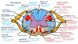
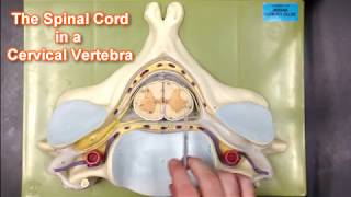
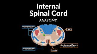
4K ultra high definition video about the spinal cord. For more information : https://www.neuromatiq.net For the spinal cord specific chapter : http://neuromatiq.net/en/chapters/2-a... The spinal cord is well protected within the spinal canal inside the spine. In adults, it measures approximately 42 cm for women and 45 cm for men. And it is at the origin of all 31 pairs of spinal nerves. The spinal cord, like the brain, is surrounded by the meninges membranes: the pia mater, the arachnoid mater and the dura mater. It's surrounded by the CSF, and has a rudimentary hole in the center (the central canal). Because of intrauterine faster growth of the spine, the nerve roots of the spinal nerves are offset relative to Inter-vertebral foramina they emerge from, That is why the spinal cord ends at the level of the second lumbar vertebra, although it gives nerves up to the fifth sacral vertebra and even the first coccyx vertebra. The Lumbar puncture for collecting CSF is usually done below the second lumbar vertebra, this prevents any injury to the spinal cord. The Spinal cord follows the path of the spine, and draws two curvatures: A Cervical with a posterior concavity (lordosis) and a dorso-lumbar with anterior concavity (kyphosis). It also has two enlargements : a cervical and a lumbar, this is due to the innervation of the upper and the lower limbs. The Spinal cord ends down with the medullary cone that gives rise to the Cauda equina (A cluster of lumbosacral nerve roots). On a cross section, the spinal cord has a central region: the gray matter which contains the neurons cell bodies, and a peripheral part: The white matter, it consists of the axonal extensions ant their myelin sheath. The gray matter has the the shape of a butterfly, with two anterior horns housing the motor neurons cell bodies and two posterior horns that receive sensory fibers. At the thoraco-lumbar level, there are also lateral horns, these hold the sympathetic fibers cell bodies. The white matter is organized into three pairs of funiculi (ventral, dorsal and lateral). The spinal cord is marked by some grooves on its surface : the deepest is the anterior median fissure (the groove in the ventral side), The posterior median sulcus is the groove in the dorsal side, The spinal cord has also two lateral grooves on each side, from which will emerge two pairs of nerve roots, a front root (for motor fibers) and the posterior root(for sensory fibers). These two nerve roots unite to form a spinal nerve on each side.
Comments