Скачать с ютуб Cervical Lymph Nodes Ultrasound Normal Vs Abnormal Images | Reactive & Malignant Neck Nodes USG Scan в хорошем качестве
Cervical Lymph Nodes Ultrasound Normal Vs Abnormal Images | Reactive & Malignant Neck Nodes USG Scan
2 месяца назад
Скачать бесплатно Cervical Lymph Nodes Ultrasound Normal Vs Abnormal Images | Reactive & Malignant Neck Nodes USG Scan в качестве 4к (2к / 1080p)
У нас вы можете посмотреть бесплатно Cervical Lymph Nodes Ultrasound Normal Vs Abnormal Images | Reactive & Malignant Neck Nodes USG Scan или скачать в максимальном доступном качестве, которое было загружено на ютуб. Для скачивания выберите вариант из формы ниже:
Загрузить музыку / рингтон Cervical Lymph Nodes Ultrasound Normal Vs Abnormal Images | Reactive & Malignant Neck Nodes USG Scan в формате MP3:
Если кнопки скачивания не
загрузились
НАЖМИТЕ ЗДЕСЬ или обновите страницу
Если возникают проблемы со скачиванием, пожалуйста напишите в поддержку по адресу внизу
страницы.
Спасибо за использование сервиса savevideohd.ru
Cervical Lymph Nodes Ultrasound Normal Vs Abnormal Images | Reactive & Malignant Neck Nodes USG Scan
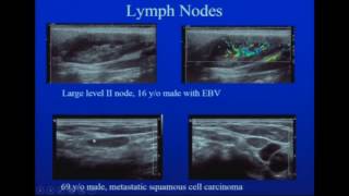
Cervical Lymph Nodes Ultrasound Normal Vs Abnormal Images | Reactive & Malignant Neck Nodes USG Scan *Cases Reactive Lymph Nodes - 0:00 Malignant Lymph Nodes - 2:36 Normal Cervical Lymph Nodes: - Shape and Size: Usually oval with a smooth, well-defined border. Measure less than 1 cm - Hypoechoic with echogenic central hilum - Vascularity: On Doppler imaging, normal lymph nodes show minimal vascularity, often with a single hilar vascular signal. Reactive Cervical Lymph Nodes: - May enlarge but usually do not exceed 2-3 cm in diameter. - Shape: They typically retain their oval shape with smooth borders, unlike the rounded shape often seen in malignant lymph nodes. - Echotexture: They have a homogeneous appearance with a preserved echogenic hilum, indicating their benign nature. - Vascularity: On Doppler ultrasound, reactive lymph nodes may show increased vascularity but it is usually centered at the hilum, which is a normal response to increased activity within the lymph node. Malignant Cervical Lymph Node: - Loss of central echogenic fatty hilum - Enlarged - Heterogeneous appearance - Become rounded or irregular in shape - Cystic necrosis: Ill-defined hypoechoic areas within lymph node - Peripheral vascularity
Comments