Скачать с ютуб Embryology of the Ear I (Easy to Understand) в хорошем качестве
Embryology of the Ear I (Easy to Understand)
3 года назад
Скачать бесплатно Embryology of the Ear I (Easy to Understand) в качестве 4к (2к / 1080p)
У нас вы можете посмотреть бесплатно Embryology of the Ear I (Easy to Understand) или скачать в максимальном доступном качестве, которое было загружено на ютуб. Для скачивания выберите вариант из формы ниже:
Загрузить музыку / рингтон Embryology of the Ear I (Easy to Understand) в формате MP3:
Если кнопки скачивания не
загрузились
НАЖМИТЕ ЗДЕСЬ или обновите страницу
Если возникают проблемы со скачиванием, пожалуйста напишите в поддержку по адресу внизу
страницы.
Спасибо за использование сервиса savevideohd.ru
Embryology of the Ear I (Easy to Understand)
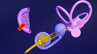
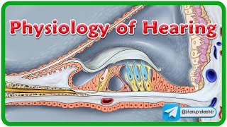
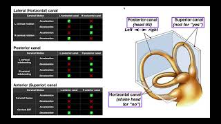
The development of the inner ear explained in 15 minutes. For the embryology of the middle and outer ear, tap here: • Embryology of the Ear II (Easy to Und... If you are completely new to embryology and you want to understand it quickly, this should be the first video you watch: - • Introduction to Embryology - Fertilis... -------------------------------- Recommended Text -------------------------------- Easy Embryology is a book that is dedicated to the simplification of embryology. It is available at https://drminass.com/product/easyembr.... Contact Dr. Minass for more information. ---------------------------------------- Interact With Dr. Minass! ---------------------------------------- Website - https://www.drminass.com Email - [email protected] Patreon - / drminass Facebook - / m1na55 Instagram - @m1.nass Post - Address to: Minass Parcel Locker 10106 04448 59 Penshurst Street Willoughby, NSW Australia 2068 ------------------------------------------------------------------------ SUMMARY OF THE VIDEO FOR YOUR NOTES ------------------------------------------------------------------------ The internal ear - The otic placode is seen in the day 22 embryo as a thickened part of the ectoderm on both sides of the rhombencephalon (developing CNS / neural tube). - the otic placode invaginates quickly forming the otic vesicles. Also known as auditory vesicles, or otocysts. - the otic placode divides itself into both a ventral component and a dorsal component - the ventral part gives rise to the saccule and the cochlear duct - the dorsal part forms the utricle, semicircular canals, and endolymphatic duct. - these are collectively the membraneous labyrinth - by week 6, the saccule has an outgrowth that eventually forms the cochlear duct by growing and coiling 2.5 times around itself. It grows into the surrounding tissue to do this. - the connection between the saccule and the cochlear duct is by the ductus reuniens - the tissue that the cochlear duct penetrated will differentiate into cartilage - some of this cartilage will become hollow, to allow for the development of the scala vestibuli and tympani, each having their own membrane. - cochlear duct is attached to the rest of the cartilage by the spiral ligament - epithelial cells of the cochlear duct differentiate - these become the outer ridge and the inner ridge - the outer ridge becomes the hair cells which are covered by the tectorial membrane - the inner ridge becomes the spiral limbus - the hair cells and the tectorial membrane are known as the Organ of Corti - the Organ of Corti sends impulses to the brain via cranial verve VIII (CN8, vestibulocochlear nerve) - by week 6 semicircular canals appear by walls that grow together - the walls disappear leaving the three semicircular canals - one of the ends become the crus ampullare that dilates and contains sensory cells, the other end doesn't dilate and is thus called the crus nonampullare - the cells in the crus amopullare are known as the crista ampullaris and these maintain equilibrium - inside the utricle and saccule , maculae acusticae develop, which send information about the bodies position in space to the brain for processing - this is done via the vestibular portion of CNVIII - the statoacoustic ganglion is formed by both neural crest cells and by some cells that divide off the optic placode (so all ectoderm) - the statoacoustic ganglion divides into both vestibular and cochlear segments (CNVIII)
Comments