Скачать с ютуб Embryology of the Eye (Easy to Understand) в хорошем качестве
Embryology of the Eye (Easy to Understand)
4 года назад
Скачать бесплатно Embryology of the Eye (Easy to Understand) в качестве 4к (2к / 1080p)
У нас вы можете посмотреть бесплатно Embryology of the Eye (Easy to Understand) или скачать в максимальном доступном качестве, которое было загружено на ютуб. Для скачивания выберите вариант из формы ниже:
Загрузить музыку / рингтон Embryology of the Eye (Easy to Understand) в формате MP3:
Если кнопки скачивания не
загрузились
НАЖМИТЕ ЗДЕСЬ или обновите страницу
Если возникают проблемы со скачиванием, пожалуйста напишите в поддержку по адресу внизу
страницы.
Спасибо за использование сервиса savevideohd.ru
Embryology of the Eye (Easy to Understand)
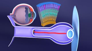
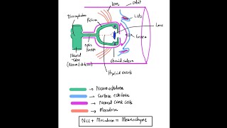
The development of the eyes explained in 15 minutes. If you are completely new to embryology and you want to understand it quickly, this should be the first video you watch: - • Introduction to Embryology - Fertilis... Post any questions you have about the video below, I read all the comments: -------------------------------- Recommended Text -------------------------------- Easy Embryology is a book that is dedicated to the simplification of embryology. It is available at https://drminass.com/product/easyembr.... Contact Dr. Minass for more information. ---------------------------------------- Interact With Dr. Minass! ---------------------------------------- Website - https://www.drminass.com Email - [email protected] Patreon - / drminass Facebook - / m1na55 Instagram - @m1.nass Post - Address to: Minass Parcel Locker 10106 04448 59 Penshurst Street Willoughby, NSW Australia 2068 ------------------------------------------------------------------------ SUMMARY OF THE VIDEO FOR YOUR NOTES ------------------------------------------------------------------------ Development of the eye is a little bit more complex than most of the organs already discussed, but as usual we will break it down simply so that we can make it easy to understand. The eye is first noticed in week three of development. Just like the development of ears are marked with the appearance of an otic placode and then an otic vesicle, the eyes are marked by the appearance of the optic placode and vesicle on either side of the embryo. As the anterior and posterior neuropores close (see CNS section), the optic placodes will form into the optic vesicles. The optic placodes are developed from neural tube (ectoderm). The optic vesicles grow until they touch the ectoderm of the skin of the embryo. The lens’ then initiate their formation, at which point the vesicle evaginates to form the optic cup. The optic cup has two layers: an inner and an outer layer. The outer layer is the pigmented layer of the retina, and the inner layer is the neural layer. In week seven, the optic cup begins to form the pupil. The actual lens itself is developed from the surface ectoderm as a lens placode, and by week five detaches itself from the surface ectoderm and is in its position inside the optic cup. The pars optica retinae is the outermost layer of the inner layer and will contain the rods and cones. Next to the pars optica retinae is the mantle layer which develops into the neurons of the eye (outer nuclear, inner nuclear, and ganglion layers). The axons of these nerves are on the surface and they join together to form the optic nerve (CNII). The most anterior portion of the inner layer has a one cell coating (pars ceca retinae) which will form both the pars iridica retinae (the inner layer of the iris) and the pars ciliaris retinae (becomes the ciliary body). The pars ciliaris retinae continually folds to give it its characteristic appearance and the ciliary muscle will eventually control the lens in response to light and other stimuli. The dilator pupillae and the sphincter of the eye form in the epithelial cells contained within the surface and the optic cup. There is another inner and outer layer for you to remember now. By week six, the tissue that surrounds the developing eye will transform into an inner and outer layer. The inner layer will become a tissue resembling the pia mater, and the outer layer resembles the dura mater. The pia mater portion differentiates into the choroid, whereas the dura mater portion will become the sclera. The anterior chamber forms when there is degeneration of the tissue that yet again forms an inner and outer layer. The inner layer is the iridopupillary membrane (which completely degenerates anterior to the lens) and the outer layer is the substantia propria. The cornea is made from the substantia propria, an epithelial layer from the ectoderm, and the epithelium shared with the anterior chamber. The posterior chamber is bordered by the iris and ciliary body. Both anterior and posterior chambers are in open communication with each other through the pupil, and both contain aqueous humor that continually circulates. Between the retina and the lens, the vitreous body is formed by infiltrating tissue that initially surrounds the optic cup.
Comments