Скачать с ютуб Renal Stone Kidney, Ureter & Bladder (KUB) Ultrasound Report Example | Renal Calculus USG Scan в хорошем качестве
Renal Stone Kidney, Ureter & Bladder (KUB) Ultrasound Report Example | Renal Calculus USG Scan
4 месяца назад
Скачать бесплатно Renal Stone Kidney, Ureter & Bladder (KUB) Ultrasound Report Example | Renal Calculus USG Scan в качестве 4к (2к / 1080p)
У нас вы можете посмотреть бесплатно Renal Stone Kidney, Ureter & Bladder (KUB) Ultrasound Report Example | Renal Calculus USG Scan или скачать в максимальном доступном качестве, которое было загружено на ютуб. Для скачивания выберите вариант из формы ниже:
Загрузить музыку / рингтон Renal Stone Kidney, Ureter & Bladder (KUB) Ultrasound Report Example | Renal Calculus USG Scan в формате MP3:
Если кнопки скачивания не
загрузились
НАЖМИТЕ ЗДЕСЬ или обновите страницу
Если возникают проблемы со скачиванием, пожалуйста напишите в поддержку по адресу внизу
страницы.
Спасибо за использование сервиса savevideohd.ru
Renal Stone Kidney, Ureter & Bladder (KUB) Ultrasound Report Example | Renal Calculus USG Scan
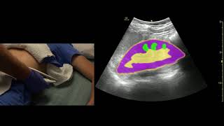
Renal Stone Kidney, Ureter & Bladder (KUB) Ultrasound Report Example | Renal Calculus USG Scan KUB Ultrasound Report: Clinical History: Patient reports persistent left flank pain for the past month, occasional nausea, and a history of kidney stones. Technique: A comprehensive ultrasound examination of the kidneys, ureters, and bladder was performed utilizing both grayscale and color Doppler imaging with a curvilinear transducer. Findings: Kidneys: The right kidney appears normal in size, shape, and parenchymal echogenicity, measuring approximately 11.2 cm in length. The left kidney is also normal in size and shape, measuring about 11 cm in length, with normal cortical thickness. However, there is a notable echogenic focus in the lower pole of the left kidney, measuring approximately 6 mm, exhibiting posterior acoustic shadowing, consistent with a renal calculus. No evidence of hydronephrosis or abnormal masses within either kidney. Ureters: The ureters are not dilated, and no stones are visualized in their course. Ureters are typically not fully visualized on ultrasound in the absence of dilation. Bladder: The bladder is adequately filled, demonstrating normal wall thickness and no intraluminal lesions. No bladder stones are seen. Post-void residual volume is within normal limits. Impression: A 6 mm renal calculus in the lower pole of the left kidney with posterior acoustic shadowing, indicative of a stone with significant density. No hydronephrosis or ureteral dilation, suggesting that the stone is currently not causing significant obstruction. Normal findings in the right kidney and bladder. Recommendations: Given the presence of a symptomatic renal stone, patient management may include increased fluid intake, pain management, and monitoring for signs of obstruction. Follow-up imaging or urology consultation may be warranted for persistent symptoms or if complications arise. Consideration for non-invasive stone removal techniques or lithotripsy if there is no spontaneous passage of the stone or in case of worsening symptoms.
Comments